Retinal Vascular Imaging & Diagnostics
In 2021, an estimated 9.6 million people in the United States had Diabetic Retinopathy (DR), with 1.8 million having vision threatening DR. Early detection and intervention can reduce a person’s risk for severe vision loss by up to 95% [1,2].
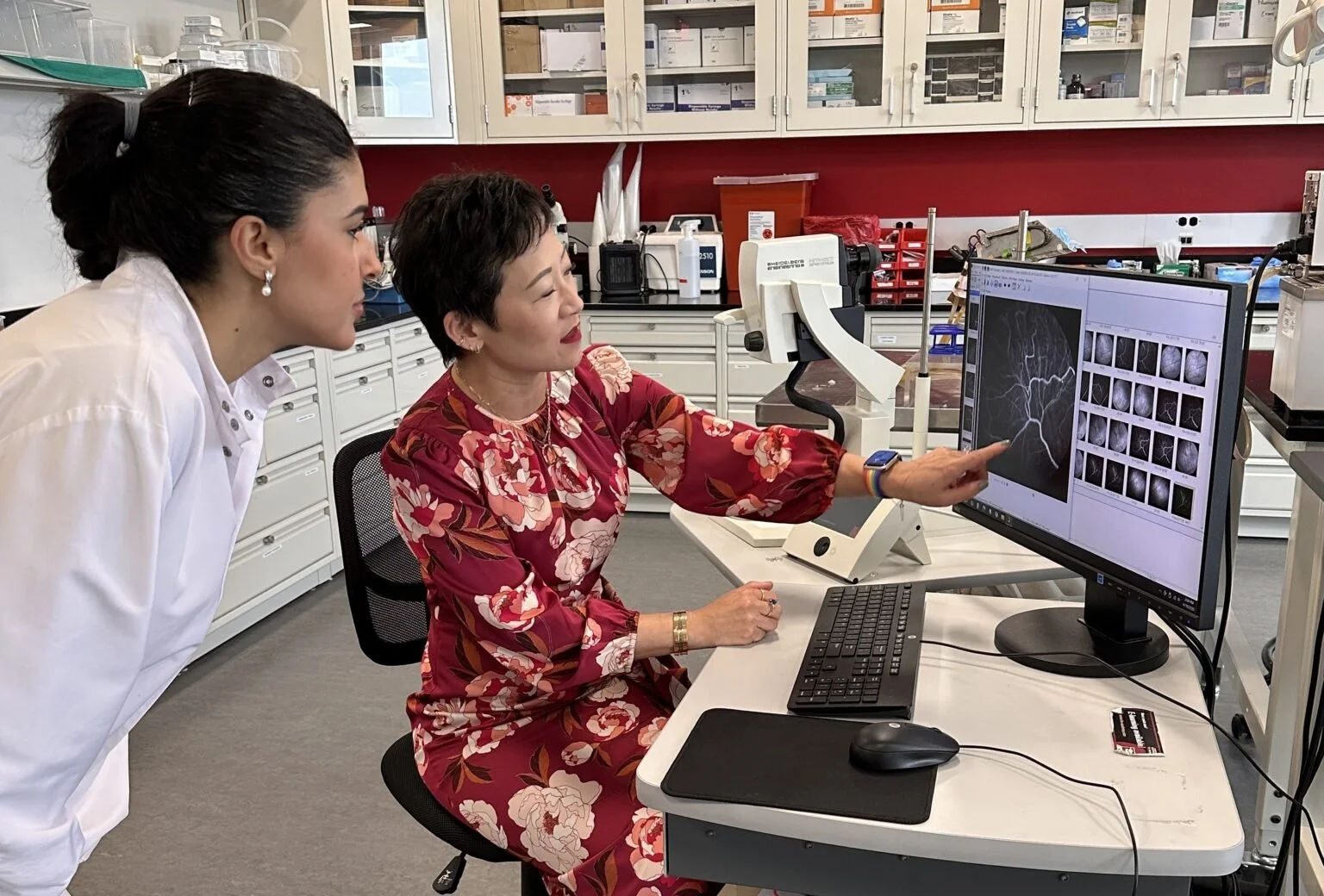
Early detection of degenerative diseases of the eye’s vasculature is essential to slowing the progression of vision loss and preserving the quality of life. Our live mammalian model lab is harnessed with advanced imaging modalities that allow researchers to view and understand the mechanism of various vascular diseases of the retina, including Diabetic Retinopathy (DR), Retinopathy of Prematurity (ROP), and Age-related Macular Degeneration (AMD).
In employing imaging diagnostics with scanning laser ophthalmoscopy (SLO), we can capture detailed retinal images, using infrared reflectance (IR), fluorescein angiography (FA), optical coherence tomography (OCT), and OCT-Angiography (OCT-A). With these advanced imaging tools, we are able to effectively visualize the intricate and complex microvasculature of the retina, deepening our understanding of disease progression to help improve existing eye health diagnostics and treatment tools.
The need for clinical eye disease detection and monitoring is increasing, and a major challenge is the widespread accessibility of high-quality diagnostic tools, particularly in rural and under-resourced areas. Leveraging smartphone camera imaging with generative AI significantly bolsters our mission of increasing access to tools that would improve early detection and intervention of rapid disease progression.
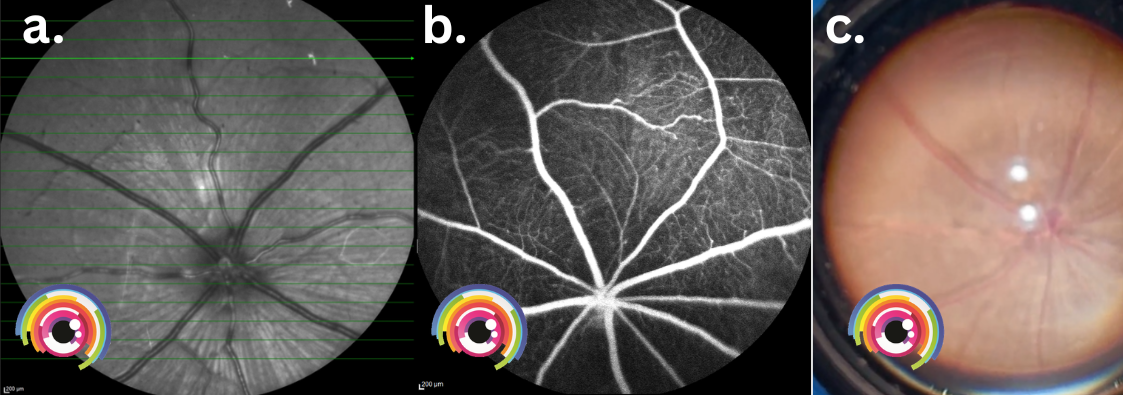
Retinal imaging using various modalities. a. IR. b. FA. c. Smartphone camera
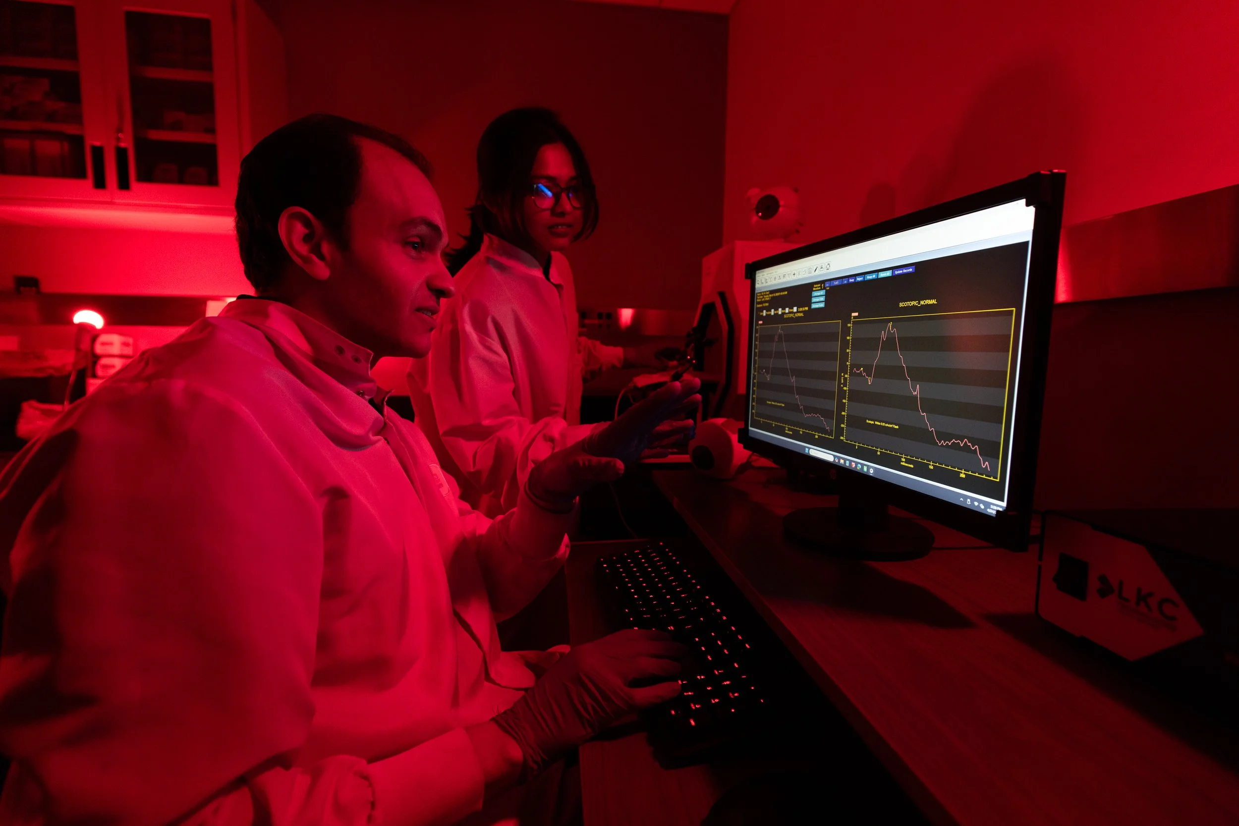
Our lab also houses a light-isolated darkroom designed specifically for electroretinography (ERG) testing. This controlled environment captures retinal responses to light stimuli that might otherwise not be captured by other avaliable imaging tools. By eliminating background light interference, we can assess retinal function under both high and low light condition, which is essential for studying visual processing of the retina and detecting ocular cellular dysfunction in a diseased state.
Vavrek, S. R., Nalbant, E. K., Konopek, N., Decker, N. L., Fawzi, A. A., Mieler, W. F., Tichauer, K. M., & Kang-Mieler, J. J. (2025). Retinal Vascular Permeability in Diabetic Subjects without Retinopathy Compared with Mild Diabetic Retinopathy and Healthy Controls. Ophthalmology Science, 5(2). https://doi.org/10.1016/j.xops.2024.100636.
Li, X., Dong, Z., Liu, H., Kang-Mieler, J. J., Ling, Y., & Gan, Y. (2023). Frequency-aware optical coherence tomography image super-resolution via conditional generative adversarial neural network. Biomedical Optics Express, 14(10), 5148–5148. https://doi.org/10.1364/boe.494557
Recent Publications
[1] https://www.cdc.gov/vision-health-data/prevalence-estimates/dr-prevalence.html
[2]https://www.nei.nih.gov/sites/default/files/2019-06/diabetes-prevent-vision-loss.pdf